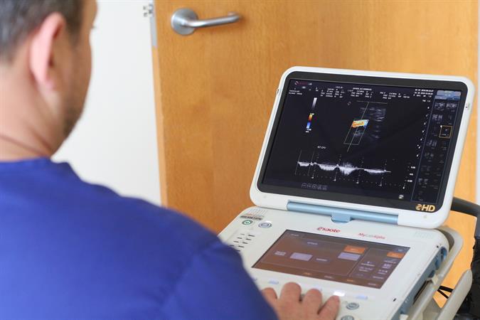
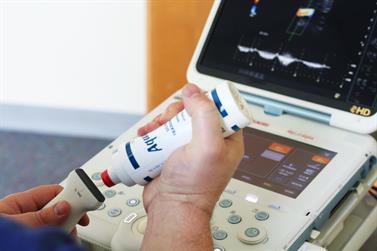
A mobile ultrasound can show how blood flows to many parts of the body. It can also tell the width of a blood vessel and reveal any blockages. These studies are a less invasive option than arteriography and venography.
Ultrasounds can help diagnose the following conditions:
- Abdominal aneurysm
- Arterial occlusion
- Blood clot
- Carotid occlusive disease
- Renal vascular disease
- Varicose veins
- Venous insufficience
We see patients in any home setting, rehab, correctional facility, as well as at industrial and OCC medical facilities for screenings.

Types of Ultrasounds
Arterial Ultrasound
What is P.A.D.?
Peripheral Arterial Disease (P.A.D.) is when plaque builds up in the arteries that carry blood to your head, limbs, and organs. P.A.D. usually affects the arteries in the legs, but can affect any major arteries in the body. This build-up of plaque is called atherosclerosis and causes a narrowing of the arteries which limits the amount of oxygenated blood to the organs and various parts of the body.
Common Symptoms
- Pain in legs while walking, known as claudication
- General pain in legs
Risk Factors
- Smoking
- Advanced Age
- Diabetes
- Hypertension (High Blood Pressure)
- Increased Cholesterol
- Coronary Heart Disease

Abdominal Ultrasound
What is an Abdominal Aneurysm?
A.A.A. (Abdominal Aortic Aneurysm) also knows as “Triple A” is a localized dilation of the abdominal aorta to twice their normal size. About 90% of these enlarged aortas form below the level of the kidney in the abdomen. A.A.A can be life threatening if it ruptures by spilling blood into the abdominal cavity. The exact reason why A.A.A. forms is still unclear; however, there are some definite risk factors.
Common Symptoms
- Pulsating sensations in abdomen
- Pain in chest, back, groin and/or abdomen
Risk factors
- Smoking (90% of all A.A.A. have occurred in people that have smoked at some point in their lives)
- Hypertension (High Blood Pressure)
- Inherited Genes
- Injury
An Ultrasound to measure the diameter of the aorta is the best way to diagnose Abdominal Aortic Aneurysm, or A.A.A.
Venous Ultrasound
What is D.V.T.?
Deep Vein Thrombosis is a condition in which a blood clot forms in one or more of the deep veins of the body. The most common place for these clots is in the legs and can occur with or without symptoms. This can become serious if the blood clot breaks loose and lodges in the lungs.
Common Symptoms
- Swelling
- Pain
- Cramping
Risk Factors
- Sitting for long periods of time
- Prolonged bed rest
- Injury or surgery
- Pregnancy
- Cancer
- Obesity
- Birth control
An ultrasound is the best way to detect blood clots in the veins.
Carotid Ultrasound
What is Carotid Artery Disease?
Carotid Artery Disease is a disease in which plaque builds up in the carotid arteries. Plaque causes the arteries to narrow. This may limit the amount of oxygenated blood to the brain. This is serious because it can lead to a stroke caused by a piece of plaque or blood clot getting stuck in a smaller artery of the brain. Carotid Artery Disease cause more than 50% of all stokes in the U.S.
Common Symptoms
- Dizziness
- Slurred speech
- Weakness to one side of the body
- Disturbed vision
- Temporary vision loss
Risk Factors
- Hypertension
- Increased cholesterol
- Diabetes
- Smoking
- Family history
The best way of detecting Carotid Artery Disease is by ultrasound.
